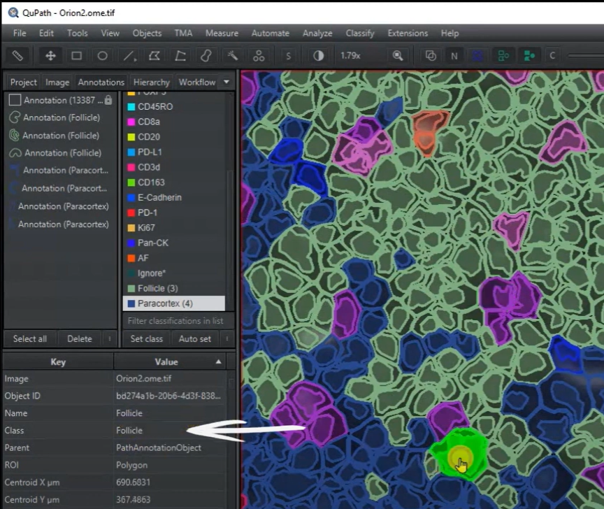
For these images I have selected creating a multiplex object classifier that looks at the proportions of various immune cells within and outside of follicles. As all of the sample images are from tonsils, there are quite a few immune cells! Other stains include some epithelial and endothelial markers, so we can also take a look at distances between vasculature and cell types. Unlike my previous walkthrough of a brightfield image analysis project, this set of videos and pages will be more exploratory and informational, to give you a handle on the tools QuPath gives you in order to analyze complex image types.
The analysis will require several steps:
Tissue outline - pixel classifier
Cell detection
Thresholder - single channel detection of areas
Cell classification - Single measurement classifier
Cell classification - Composite classifier
Pixel classification - detecting complex regions like follicles
??? Mix and match all of your tools!
In order to set you up for success, however, we will start by taking a step back and building a project, discussing what a project is, and then exploring some of the visualization options.
